Introduction to Medical Image Segmentation
Understanding Medical Image Segmentation
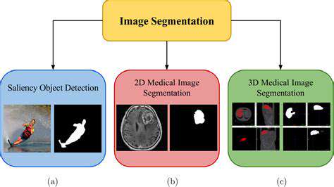
Medical image segmentation is a crucial aspect of medical image analysis, playing a vital role in various applications, from disease diagnosis to treatment planning. It involves the process of partitioning a medical image into meaningful regions or segments based on their characteristics. This process often relies on sophisticated algorithms to accurately identify and delineate anatomical structures, tissues, or abnormalities within the image, such as tumors, organs, or blood vessels. The accuracy of the segmentation directly impacts the reliability of subsequent analyses and decisions made by healthcare professionals.
The goal is to precisely delineate specific structures within the image, allowing for detailed quantitative analysis and ultimately leading to better clinical outcomes. This meticulous process is essential for tasks like measuring organ volumes, assessing tissue composition, and detecting subtle changes indicative of disease. Successful segmentation requires algorithms that can robustly identify and classify diverse tissue types, often with varying intensities and textures.
Types of Medical Images Used
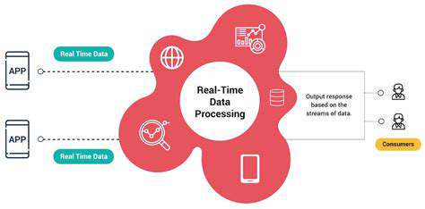
Medical image segmentation operates on a variety of imaging modalities, each with its unique characteristics and challenges. CT scans, for instance, provide detailed cross-sectional views of the body, offering high resolution and contrast for visualizing bone structures and internal organs. MRI scans, on the other hand, excel at visualizing soft tissues, providing superior contrast and detail for identifying subtle abnormalities and lesions. Ultrasound images, known for their real-time capabilities, are particularly useful for assessing moving structures like the heart and blood vessels. Each modality presents specific challenges in terms of image quality, noise levels, and contrast variations, demanding tailored segmentation algorithms to achieve accurate results.
Different imaging techniques, each with their own strengths and weaknesses, necessitate the development of customized segmentation methods to effectively extract and analyze the desired information, ultimately benefiting diagnostic and therapeutic procedures.
Challenges in Medical Image Segmentation
Accurate medical image segmentation presents a multitude of challenges. Variations in image quality, including noise, artifacts, and varying intensities, can significantly impact segmentation accuracy. Furthermore, the intricate anatomical structures and diverse tissue types found in medical images can be difficult for algorithms to distinguish. The complexity of the underlying anatomical features and the variability in tissue characteristics require robust and adaptable segmentation algorithms to accurately and reliably delineate different regions of interest.
Applications of Medical Image Segmentation
Medical image segmentation finds widespread applications in various medical fields. In oncology, it aids in tumor detection and volume measurement, enabling more accurate staging and treatment planning. In cardiology, it supports the assessment of cardiac function and the identification of abnormalities in blood vessels. In neurology, it aids in the analysis of brain structures and the detection of neurological disorders. These applications highlight the crucial role of accurate segmentation in providing valuable insights into disease characteristics and facilitating informed clinical decisions.
Beyond these specific examples, segmentation plays a critical role in various other medical applications, driving advancements in diagnostics, treatment planning, and overall patient care.
Role of Artificial Intelligence (AI)
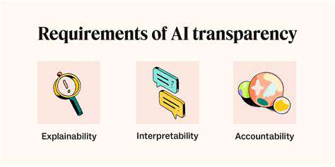
Artificial intelligence (AI), specifically deep learning, has revolutionized medical image segmentation. Deep learning models, with their ability to learn complex patterns from vast amounts of data, excel at segmenting diverse anatomical structures and tissues with remarkable precision. These models can be trained on large datasets of annotated medical images, enabling them to identify subtle features and nuances that might be missed by traditional methods. This capability leads to more accurate and reliable segmentation results, which are vital for improving the efficiency and effectiveness of medical diagnoses and treatments.
The integration of AI into medical image segmentation has led to significant improvements in accuracy and efficiency, paving the way for faster and more precise diagnostic processes.
Future Directions and Conclusion
The future of medical image segmentation holds immense promise, driven by advancements in AI and deep learning. Researchers are actively exploring novel techniques to enhance the robustness and adaptability of segmentation algorithms, particularly in addressing the challenges posed by diverse imaging modalities and variations in image quality. The development of more sophisticated and efficient AI models, combined with larger and more diverse datasets, will continue to improve the precision and reliability of segmentation results. This ongoing advancement promises to further enhance the accuracy of disease diagnosis, treatment planning, and ultimately, patient care.
In conclusion, medical image segmentation, with its integration of AI, holds a pivotal role in shaping the future of healthcare.